This site is supported by our readers. We may earn a commission, at no cost to you, if you purchase through links.
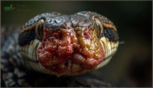
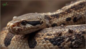
When a timber rattlesnake was found in New Hampshire with its face marred by crusty lesions and clouded eyes, veterinarians initially suspected trauma or bacterial infection. The culprit turned out to be far more insidious: Ophidiomyces ophiodiicola, a specialized fungus that has quietly invaded snake populations across at least 23 states since 2006.
This pathogen doesn’t just create superficial wounds—it breaches the skin barrier, triggering lesions at every abraded site and sometimes advancing into the eyes, throat, and lungs.
Identifying snake fungal disease early requires understanding both the visible markers and the diagnostic tools that separate it from similar conditions, because in threatened species already struggling with habitat loss, a 66.5% infection rate can tip the balance toward local extinction.
Table Of Contents
Key Takeaways
- Snake Fungal Disease, caused by Ophidiomyces ophiodiicola, has spread across 23+ U.S. states since 2006 and now threatens 42+ snake species with infection rates reaching 66.5% in some populations, making early detection critical for conservation efforts.
- You can identify SFD through visible markers including crusted facial lesions, abnormal shedding patterns (dysecdysis), skin discoloration on ventral surfaces, and behavioral changes like prolonged basking and reduced activity—symptoms that progress internally to eyes, throat, and lungs if untreated.
- Diagnosis requires laboratory confirmation through histopathology to detect fungal hyphae and PCR testing showing 74-86% accuracy, since visual symptoms alone can’t definitively separate SFD from other skin conditions affecting snakes.
- Prevention depends on strict biosecurity protocols including 30-day quarantine periods, 3% bleach disinfection of equipment, humidity control below 60%, and when infection occurs, terbinafine nebulization treatment shows clinical improvement within 58-110 days though severe cases carry 66.7% mortality rates.
What is Snake Fungal Disease?
Snake Fungal Disease is an emerging wildlife health threat that’s reshaping how we comprehend reptile diseases in North America and beyond. The culprit behind this illness is Ophidiomyces ophiodiicola, a specialized fungus that targets snake species with sometimes devastating results.
To recognize and respond to this disease effectively, you need to understand what causes it, how it spread across the continent, and which snakes face the greatest risk.
Overview of Ophidiomyces Ophiodiicola
Snake fungal disease stems from a specialized fungus with distinct preferences. Ophidiomyces ophiodiicola, the culprit behind this devastating condition, belongs to the family Onygenaceae and thrives under surprisingly specific conditions that shape its infection process. The disease was first observed in timber rattlesnakes in 2006.
- Fungal taxonomy: Reclassified in 2013 from Chrysosporium to Ophidiomyces within order Onygenales
- Growth conditions: Prefers alkaline environments (pH 9) and fails above 95°F
- Infection process: Breaches snake skin, causing 100% lesion development at abraded sites
- Geographic spread: Detected across 23 US states and confirmed in 33 snake species
- Transmission routes: Environmental substrates and direct contact promote fungal disease spread
History and Spread of SFD
Understanding the fungus is one thing, but tracking its march across continents reveals the scope of this wildlife disease threat. Snake Fungal Disease first surfaced in New Hampshire during 2006, though retrospective evidence traces it to 1945 in North America and 1959 in Europe.
Geographic expansion accelerated dramatically after 2008, with disease surveillance now confirming infections across 30+ U.S. states, Canada, and multiple European nations. The disease was recently confirmed when wild snakes tested positive in Europe.
Environmental contamination and disease transmission through both wild and captive populations drove these global outbreaks, transforming a localized concern into an international conservation crisis.
Snake Species Commonly Affected
This fungal pathogen doesn’t discriminate. Over 42 wild snake species across three continents have tested positive for Ophidiomyces ophiodiicola as of 2024.
Species susceptibility spans six snake families—from colubrids to vipers—with prevalence rates ranging from 1.8% to 66.5% depending on geographic distribution.
Threatened species like the Texas-listed Nerodia harteri harteri face 50% infection rates, amplifying conservation impact where populations already struggle.
Threatened snake species face staggering 50% infection rates, turning a fungal disease into a conservation crisis for already vulnerable populations
Recognizing Early Signs of SFD
Catching snake fungal disease early can mean the difference between recovery and decline. You’ll notice changes in your snake’s appearance and behavior before the infection takes hold internally.
Here’s what to watch for in the early stages of SFD.
Visible Skin Lesions and Discoloration
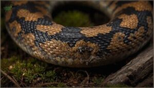
One of the earliest warning signs you’ll notice is subtle changes in your snake’s skin—discolored scales, crusting, or erosion along scale margins. Lesion prevalence varies widely, with studies reporting 14.5% to 44% of wild populations affected.
Most skin lesions appear on the ventral surface (96.9%), where contact with contaminated substrate occurs. Fungal confirmation through PCR testing reveals Ophidiomyces ophiodiicola DNA in 74-86% of sampled lesions, establishing the diagnostic link between visible dermatitis and Snake Fungal Disease symptoms.
Unusual Shedding Patterns (Dysecdysis)
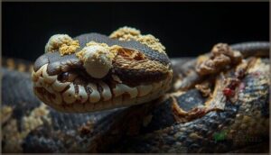
When your snake sheds in ragged patches instead of one smooth piece, you’re witnessing dysecdysis—a telltale Snake Fungal Disease symptom. Retained eye caps appear cloudy and adherent, while skin thickening creates yellowish crusts around the head.
Incomplete shedding leaves old skin stuck to limbs and tail, with skin adherence causing painful tearing. These unusual shedding patterns, combined with skin lesions and ulceration, demand immediate Snake Fungal Disease diagnosis.
Facial Swelling and Disfigurement
You’ll spot facial swelling—the most reliable clinical sign—in nearly every late-stage snake fungal infection case. This disfigurement progresses from mild puffiness to severe distortion, with Snake Fungal Disease diagnosis confirmed through histopathology findings revealing fungal hyphae penetrating facial tissues.
Swelling severity correlates directly with fungal burden, making monitoring trends essential for diagnostic accuracy across species susceptibility ranges.
Behavioral Changes in Infected Snakes
When your snake stops eating or stays exposed longer than usual, you’re witnessing reduced activity that signals snake fungal disease. Infected snakes show basking alterations—spending twice as long in the open—alongside foraging disruption and impaired predator avoidance.
These snake fungal infection symptoms reflect energetic cost: lethargy, weight loss, and delayed responses. Snake fungal disease impacts become undeniable when movement drops 35% below baseline rates.
Advanced Symptoms and Disease Progression
Once SFD takes hold, the infection doesn’t stay on the surface—it moves deeper into a snake’s body, affecting organs and systems that are critical for survival. Understanding how the disease progresses helps you recognize when a snake has moved past the early stages and needs immediate intervention.
You’ll see the external damage worsen with spreading lesions and nodules, but the internal toll often proves more dangerous.
Internal Involvement (Eyes, Throat, Lungs)
When snake fungal disease advances, you’ll notice the infection doesn’t stay on the surface. Internal dissemination patterns reveal systemic infection spreading to key organs, creating life-threatening complications:
- Ocular manifestations – cloudy eyes, corneal opacity, and periocular swelling appear in up to 15% of later cases
- Throat lesions – ulcerative stomatitis affecting nearly 10% of infected wild snakes
- Pulmonary involvement – respiratory distress from lung tissue nodules, contributing to 40% mortality rates
Experimental insights confirm snake fungal infection symptoms progress internally within 30–37 days.
Development of Nodules and Ulcerations
As infection deepens, you’ll observe abnormal subcutaneous bumps—nodules—forming within 12 to 30 days post-exposure. Ulcerations, full-thickness skin erosions affecting up to 7.7% of wild snakes, signal severe progression. Lesion distribution favors the ventral surface in 96.9% of cases, with nodule formation often preceding ulceration.
Diagnostic confirmation requires histopathology and qPCR to detect fungal hyphae within granulomatous tissue.
Respiratory Issues and Lethargy
When Ophidiomycosis advances, you’ll notice respiratory distress—open-mouth breathing, wheezing, or nasal discharge—affecting roughly 10–15% of cases during late-stage disease progression.
Pulmonary invasion damages lung tissue, with experimental mortality exceeding 40% when systemic complications develop.
Lethargy prevalence reaches 70% in severe snake fungal infection symptoms, marked by prolonged immobility, reduced feeding, and abnormal basking. These physiological sequelae signal critical internal involvement requiring immediate intervention.
Diagnostic Methods for Snake Fungal Disease
When you suspect snake fungal disease, getting the right diagnosis separates guesswork from certainty. Visual symptoms alone won’t confirm Ophidiomyces ophiodiicola, so veterinarians rely on specific laboratory techniques to pinpoint the infection.
Here’s how professionals identify SFD with accuracy.
Physical Examination and Clinical Signs
Diagnosis starts with systematic observation of external features and behavioral indicators. You’ll want to examine your snake for visible abnormalities that signal infection. Key physical findings include:
- Lesion morphology: crusted, ulcerated, or thickened areas on scales
- Scale abnormalities: discoloration, erosion at margins, nodules beneath skin
- Facial swelling: disfigurement impairing feeding ability
- Unusual shedding: incomplete or accelerated shed cycles
- Behavioral indicators: altered basking routines, reduced appetite
Skin lesions consistent with snake fungal infection symptoms appear in roughly 25% of surveyed wild populations.
Histopathology and Microscopic Analysis
While physical signs point toward infection, laboratory confirmation is where the real diagnostic power lies. Tissue samples undergo microscopic examination to reveal fungal elements—branching hyphae measuring 1–4 μm in diameter—invading deeper skin layers.
Pathologists identify granulomatous dermatitis with necrosis in nearly all confirmed cases, grading lesion severity through standardized scoring systems. This histopathological analysis definitively separates SFD from similar skin conditions.
Preventing and Managing SFD in Snakes
Once you’ve identified SFD in a snake, the next challenge is preventing its spread and giving the infected animal the best chance at recovery. Management strategies differ depending on whether you’re working with captive or wild populations, but the principles remain consistent across settings.
The following sections outline practical approaches to reduce transmission, explore available treatment options, and implement supportive care measures that can improve outcomes.
Reducing Transmission in Captivity and Wild
To break the cycle of disease transmission and prevention, you’ll need a multi-layered approach combining captive quarantine, biosecurity measures, and field protocols.
Isolate newly acquired snakes for 30 days—this prevents introducing Ophidiomyces into your collection. Disinfect boots and equipment with 3% bleach between handling events.
Environmental management, including humidity control below 60%, reduces fungal proliferation by 44%.
Population monitoring through regular PCR screening catches asymptomatic carriers early, protecting both captive and wild snake populations.
Treatment Options and Their Effectiveness
Once you’ve identified SFD, antifungal drug therapy becomes your primary weapon. Terbinafine nebulization shows marked clinical improvement—12 Lake Erie water snakes cleared visible lesions over 110 days with no adverse effects. PCR-negative swabs appeared after 58 days of treatment.
However, case fatality rates reach 66.7% in severe cases, making early intervention critical. Treatment duration averages eight weeks, with nebulized delivery outperforming oral routes in comparative effectiveness studies.
Environmental and Supportive Care Strategies
Beyond pharmaceuticals, you’ll need thorough environmental contamination and control measures. Maintain strict biosecurity protocols—disinfect equipment with 3% bleach for a minimum of two minutes. Implement quarantine isolation for at least 30 days.
Humidity control proves essential, as moisture accelerates fungal survival. Provide fluid management through fresh water ad libitum while carefully titrating nutritional support—overfeeding gravid females increases reproductive failures by 80%.
These reptile health and wellness strategies complement veterinary care for snakes during fungal infection treatment.
Frequently Asked Questions (FAQs)
Can humans contract Snake Fungal Disease from snakes?
No confirmed human infections exist. You can’t contract SFD—Ophidiomyces ophiodiicola lacks zoonotic potential, showing host specificity to reptiles.
However, you might carry fungal spores on clothing, indirectly transmitting the pathogen between snakes.
How long can the fungus survive outdoors?
Ophidiomyces ophiodiicola persists in contaminated soil for at least 30 days under laboratory conditions, with temperature tolerance, moisture influence, and seasonal variation affecting fungal pathogens’ viability in outdoor habitats year-round.
Are there vaccines available for snake populations?
While research into mammalian fungal vaccines continues, no approved vaccines exist for snakes battling Ophidiomyces ophiodiicola.
You’ll rely on antifungal medications, PCR testing, and biosecurity measures until future vaccine prospects materialize.
Does climate change influence SFD spread patterns?
Yes, climate change influences SFD spread. Temperature effects and moisture influence pathogen growth, while habitat changes stress host immunity.
Future projections show warming winters may increase disease severity, threatening ecosystem health and wildlife populations.
What disinfection methods kill Ophidiomyces ophiodiicola effectively?
You’ll need effective disinfectants to eliminate Ophidiomyces ophiodiicola and prevent fungal infection spread. Bleach disinfection at 3–10% concentration for two minutes achieves complete fungal eradication, while quaternary ammonia products and hydrogen peroxide combinations also demonstrate strong disinfectant efficacy.
These methods are integral to antifungal treatment protocols for reptile disease diagnosis and treatment and animal disease management and control.
Conclusion
Studies show that 66.5% of timber rattlesnakes in certain populations carry active infections, underscoring how rapidly this pathogen can compromise vulnerable species.
Your ability to distinguish snake fungal disease identification guide criteria—from crusty facial lesions to abnormal shedding—directly influences whether conservation efforts succeed or fail.
Early detection through systematic observation and histopathology remains the most reliable defense, turning what might become a lethal epidemic into a manageable threat with prompt intervention and targeted care protocols.
- https://pmc.ncbi.nlm.nih.gov/articles/PMC7544097/
- https://www.nature.com/articles/s41598-024-55354-5
- https://www.usgs.gov/publications/prevalence-ophidiomyces-ophidiicola-and-epizootiology-snake-fungal-disease-free
- https://www.in.gov/dnr/fish-and-wildlife/wildlife-resources/animals/snakes/snake-fungal-disease/
- https://www.frontiersin.org/journals/veterinary-science/articles/10.3389/fvets.2021.665805/full
