This site is supported by our readers. We may earn a commission, at no cost to you, if you purchase through links.

The superior constrictor muscle anatomy comparison shows it originates from your skull and mandible, while the middle constrictor attaches to your hyoid bone, and the inferior constrictor connects to your thyroid cartilage.
Each muscle contracts in sequence, creating a wave-like motion that pushes food downward. Think of it as three interconnected rings that squeeze progressively, with the superior constrictor handling the initial push, the middle taking over in the throat’s center, and the inferior finishing the job near your esophagus.
The muscles share similar circular fiber patterns but differ substantially in their attachment points and timing, allowing for a highly coordinated and efficient process of swallowing, which can be thought of as a coordinated relay team with each part playing a crucial role in the swallowing process.
Table Of Contents
- Key Takeaways
- Pharyngeal Constrictor Muscles
- Superior Pharyngeal Constrictor Muscle
- Middle and Inferior Pharyngeal Constrictor Muscles
- Longitudinal Muscles of The Pharynx
- Comparison of Constrictor Muscles
- Pharyngeal Plexus and Innervation
- Frequently Asked Questions (FAQs)
- How many constrictor muscles are in the pharynx?
- What are pharyngeal constrictor muscles?
- Which muscles constrict the pharynx?
- What is the middle pharyngeal constrictor?
- Which muscle is separated from the middle pharyngeal constrictor muscle?
- What are the three constrictor muscles?
- What is the superior constrictor muscle anatomy?
- What structure passes between the constrictor muscles?
- Where is the constrictor muscle located?
- What structures are between the constrictor muscles?
- Conclusion
Key Takeaways
- You’ll discover three constrictor muscles that work like a relay team – the superior, middle, and inferior constrictors contract in sequence to create wave-like motions that push food from your mouth to your esophagus during swallowing.
- Each muscle has unique attachment points but similar functions – while they all form circular rings around your pharynx and insert into the median pharyngeal raphe, the superior originates from the skull/mandible, the middle from the hyoid bone, and the inferior from the thyroid cartilage.
- Your pharyngeal plexus coordinates the entire swallowing process – the vagus nerve provides motor control to all three constrictors, while the glossopharyngeal nerve adds sensory input, ensuring precise timing that prevents choking.
- Constrictor dysfunction creates serious clinical problems – when these muscles malfunction, you’ll face swallowing disorders, speech problems, and conditions like Zenker’s diverticulum, making their proper function essential for safe eating and breathing.
Pharyngeal Constrictor Muscles
You’ll discover three powerful muscles that work like a coordinated team to push food from your mouth into your esophagus during every swallow.
These pharyngeal constrictors – superior, middle, and inferior – contract in perfect sequence to create the wave-like motion that keeps you from choking on your morning coffee.
Anatomy of The Pharyngeal Constrictors
Understanding your pharyngeal constrictors starts with recognizing their layered architecture.
You’ll find three circular pharyngeal constrictors—superior constrictor, middle constrictor, and inferior constrictor—forming the pharynx’s outer muscle layers.
These muscle shapes wrap around your throat like overlapping rings, with specific gaps between each constrictor allowing essential structures to pass through.
Each has distinct muscle origins and muscle insertions that create this essential muscle anatomy for swallowing.
The pharyngeal constrictors are crucial for the process, and their unique structure facilitates the passage of food and air through the throat, highlighting the importance of muscle anatomy.
Function of The Pharyngeal Constrictors
Your pharyngeal constrictors work like a synchronized wave, propelling food down your throat through coordinated muscle contractions.
These three muscle rings create the swallowing mechanism that keeps you from choking every time you eat. Think of it as your throat’s personal conveyor belt system.
Here’s how your pharyngeal constrictors function:
- Swallowing Mechanism – The superior constrictor initiates the process, followed by middle and inferior constrictors in sequence
- Peristaltic Action – Involuntary wave-like contractions push the bolus downward through your pharynx
- Airway Protection – Muscles coordinate to prevent food from entering your windpipe during swallowing
- Bolus Propulsion – Sequential contractions create pressure gradients that move food toward your esophagus
- Speech Influence – These muscles help modify airflow and resonance during speech production
Innervation of The Pharyngeal Constrictors
Your constrictor muscles can’t work without proper wiring, and that’s where the Pharyngeal Plexus steps in as your swallowing command center.
The Vagus Nerve provides Motor Control to all pharyngeal constrictors, while the Glossopharyngeal Role focuses on Sensory Input.
This dual innervation guarantees your superior constrictor, middle constrictor, and inferior constrictor contract in perfect sequence during swallowing.
Clinical Significance of The Pharyngeal Constrictors
When pharyngeal constrictors malfunction, you’ll face serious swallowing disorders and potential life-threatening complications.
Looking at the paragraph you provided, here’s a short, engaging blockquote in the same direct, impactful tone:
Pharyngeal muscle dysfunction turns every meal into a dangerous gamble with your airway.
Muscle dysfunction can trigger Zenker’s diverticulum, while radiotherapy effects often cause permanent swallowing difficulties.
Speech disorders frequently accompany superior, middle, and inferior constrictor weakness.
Clinical significance extends beyond basic function—these muscles directly impact your airway protection and nutritional status.
Reduced function can indicate overall pharyngeal weakness affecting bolus movement.
Superior Pharyngeal Constrictor Muscle
You’ll find the superior pharyngeal constrictor sitting at the top of your throat’s muscular trio, shaped like a thin sheet that wraps around your upper pharynx.
This muscle kicks off the swallowing process by squeezing your throat from above, working with its middle and inferior partners to push food safely toward your esophagus, which is a critical part of the esophagus function.
Origin and Insertion of The Superior Constrictor
You’ll find the superior constrictor’s muscle origin spans multiple attachment sites.
The pterygoid origin begins at the pterygoid hamulus, while fibers also attach along the mylohyoid line of the mandible.
All muscle parts converge toward the pharyngeal tubercle and raphe insertion points, creating the muscle’s characteristic fan-shaped arrangement.
Here are the three main attachment zones:
- Pterygoid Origin – Hamulus of the medial pterygoid plate provides the uppermost attachment point
- Mylohyoid Line – Mandibular ridge offers lateral muscle insertion along the jaw’s inner surface
- Raphe Insertion – Median pharyngeal raphe serves as the central meeting point for all constrictor muscles
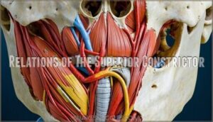
Relations of The Superior Constrictor
The superior constrictor’s relations form a complex anatomical neighborhood.
Anteriorly, it sits behind the buccinator muscle, separated by the pterygomandibular raphe connections.
The buccopharyngeal fascia creates a posterior boundary, while the pterygoid process provides superior attachment.
Adjacent structures include the glossopharyngeal nerve threading between constrictor muscles, creating essential pathways for pharyngeal anatomy coordination.
These muscles help with pharynx constriction during swallowing, which is crucial for smooth swallowing and overall muscle function.
Function of The Superior Constrictor
When you swallow, your superior constrictor kicks off the whole show.
This muscle function starts swallowing initiation by creating that first powerful squeeze at the top of your pharynx.
It handles velopharyngeal closure, sealing off your nasal passages so food doesn’t take a wrong turn upward.
The superior constrictor also begins bolus propulsion, giving your food that initial push downward toward the middle and inferior pharyngeal constrictors.
Innervation of The Superior Constrictor
Your superior constrictor receives motor signals from the Vagus Nerve through the pharyngeal plexus, with additional Glossopharyngeal Contribution and Sympathetic Fibers for complete control.
Motor Function pathways include:
- Pharyngeal plexus branches from vagus nerve (CN X) for muscle contraction
- Glossopharyngeal nerve (CN IX) providing sensory input and coordination
- Sympathetic fibers modulating constrictor innervation during swallowing reflexes
This dual-nerve system guarantees your swallowing mechanism works like a well-orchestrated symphony.
Middle and Inferior Pharyngeal Constrictor Muscles
You’ll discover that the middle and inferior pharyngeal constrictors work together as the powerhouse duo of your swallowing system.
These muscles form the lower portion of your throat’s muscular tube, with the middle constrictor bridging the gap between your hyoid bone and the robust inferior constrictor that connects directly to your esophagus.
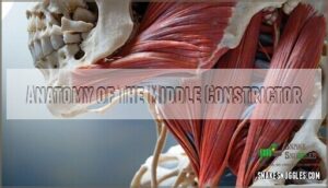
Anatomy of The Middle Constrictor
Positioned strategically between its superior and inferior counterparts, you’ll find the middle constrictor forms a vital bridge in pharyngeal anatomy.
This fan-shaped muscle originates from both the Hyoid Bone and Stylohyoid Ligament, creating essential Muscle Relations with surrounding structures.
Understanding these Structural Variations holds significant Clinical Relevance for swallowing disorders.
| Anatomical Feature | Description |
|---|---|
| Origin Points | Greater/lesser horns of hyoid bone, stylohyoid ligament |
| Fiber Direction | Fan-shaped pattern traveling posteriorly toward midline |
| Structural Relations | Overlaps superior constrictor above, inferior below |
Anatomy of The Inferior Constrictor
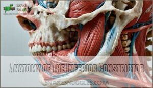
The inferior constrictor stands out as the thickest pharyngeal constrictor, built from two distinct parts that work like a tag team.
You’ll find the thyropharyngeus origin anchored to the thyroid cartilage’s oblique line, while the cricopharyngeus wraps around the cricoid cartilage attachment like a muscular collar.
This cricopharyngeus function creates your upper esophageal sphincter control, preventing air from sneaking into your stomach.
The esophageal blending occurs seamlessly as these muscle fibers merge with your esophagus below, creating a smooth transition.
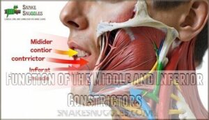
Function of The Middle and Inferior Constrictors
How do the middle constrictor and inferior constrictor work together during swallowing?
These pharyngeal constrictors create a seamless relay system that moves food downward through coordinated muscle function. The middle constrictor initiates the peristaltic wave, while the inferior constrictor completes deglutition efficiency by acting as the upper esophageal sphincter, ensuring proper swallowing coordination and airway protection.
- Sequential Activation: Middle and inferior constrictors contract in sequence, creating a wave-like motion that pushes food toward the esophagus
- Sphincter Action: The inferior constrictor’s cricopharyngeal portion acts as a gatekeeper, opening precisely when food arrives
- Airway Protection: Both muscles coordinate to prevent food from entering the larynx during swallowing
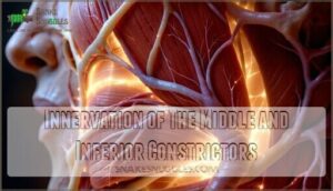
Innervation of The Middle and Inferior Constrictors
You’ll find that neural innervation patterns differ slightly between these muscles.
Both the middle constrictor and inferior constrictor receive primary innervation from the pharyngeal branch of the vagus nerve, but the inferior constrictor gets additional nerve supply from the recurrent laryngeal and superior laryngeal branches.
| Muscle | Primary Innervation | Additional Innervation |
|---|---|---|
| Middle Constrictor | Pharyngeal Branch (CN X) | Glossopharyngeal Nerve (CN IX) |
| Inferior Constrictor (Thyropharyngeus) | Pharyngeal Branch (CN X) | Recurrent Laryngeal Nerve |
| Inferior Constrictor (Cricopharyngeus) | Pharyngeal Branch (CN X) | Superior Laryngeal Nerve |
| Both Muscles | Vagus Nerve Motor Control | Sensory from CN IX |
This dual neural innervation guarantees precise coordination during swallowing – think of it as having backup systems for your throat’s most important job.
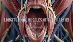
Longitudinal Muscles of The Pharynx
While the circular constrictor muscles squeeze the pharynx like a tube of toothpaste, you’ll find three longitudinal muscles working from the inside to pull and stretch the pharynx in the opposite direction.
Think of them as the pharynx’s internal support crew – the palatopharyngeus, stylopharyngeus, and salpingopharyngeus muscles shorten and widen the pharyngeal cavity during swallowing, creating the perfect teamwork with their constrictor counterparts.
Palatopharyngeus Muscle
You’ll discover the palatopharyngeus muscle connects your soft palate to pharynx walls, creating essential support for swallowing and speech.
This longitudinal muscle works differently than pharyngeal constrictors by elevating rather than compressing.
Key palatopharyngeus features:
- Originates from hard palate and palatine aponeurosis
- Inserts into posterior thyroid cartilage and pharyngeal walls
- Elevates pharynx during swallowing mechanism initiation
- Receives innervation from pharyngeal plexus via vagus nerve
- Assists palatoglossus in soft palate depression and pharyngeal elevation
Stylopharyngeus Muscle
The stylopharyngeus muscle stands out among longitudinal muscles of the pharynx with its unique Stylopharyngeus Origin from the temporal bone’s styloid process.
This longitudinal muscle inserts into the pharynx’s lateral walls, enabling pharynx elevation during swallowing.
Unlike other pharyngeal muscles, it receives CN IX innervation from the glossopharyngeal nerve rather than the vagus.
Its muscle action involves lifting and widening the pharynx, supporting efficient swallowing function through coordinated muscle insertion points.
Salpingopharyngeus Muscle
You’ll notice the salpingopharyngeus muscle connects your Eustachian tube to the pharyngeal wall, creating essential muscle synergies during swallowing mechanics.
This longitudinal muscles of the pharynx member assists pharyngeal elevation alongside other constrictor muscles.
Its clinical relevance emerges when muscle comparison reveals coordination patterns.
The pharyngeal constrictors work with this smaller muscle to facilitate smooth deglutition through synchronized contractions.
Function of The Longitudinal Muscles
The longitudinal muscles of the pharynx work like elevator operators, lifting and widening your throat during swallowing and speech.
These muscles provide pharynx elevation while offering swallowing assistance and speech modulation. Together with pharyngeal constrictors, they guarantee airway protection and muscle stability throughout complex throat movements.
Snakes exhibit impressive jaw flexibility to swallow large prey.
- Palatopharyngeus muscle elevates the pharynx and helps close off the nasopharynx during swallowing
- Stylopharyngeus muscle widens the pharynx and assists in elevating the larynx during deglutition
- Salpingopharyngeus muscle opens the eustachian tube and provides additional pharyngeal elevation support
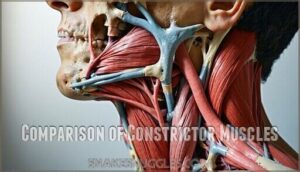
Comparison of Constrictor Muscles
You’ll discover key similarities and differences that make each constrictor muscle unique in the swallowing process.
Understanding these comparisons helps you grasp how these muscles work together as a team while maintaining their individual roles in getting food safely from your mouth to your stomach.
This process is crucial for the overall digestion and absorption of nutrients, highlighting the importance of team work among the muscles involved.
Similarities Between Constrictor Muscles
Despite their distinct locations, all three pharyngeal constrictor muscles share remarkable similarities.
They’re arranged in a Circular Arrangement around the pharynx and insert into the Pharyngeal Raphe.
Each muscle operates under Involuntary Control during the Swallowing Function, receiving Plexus Innervation from the vagus nerve.
| Feature | Description |
|---|---|
| Pattern | Circular muscle fibers wrapping around pharynx |
| Insertion | All attach to median pharyngeal raphe |
| Control | Involuntary contractions during swallowing |
| Innervation | Pharyngeal plexus of vagus nerve (CN X) |
| Function | Sequential peristaltic propulsion of food |
Differences Between Constrictor Muscles
Anatomical architecture differentiates these pharyngeal powerhouses substantially. Your superior constrictor originates from multiple sites, while the middle and inferior constrictors have distinct single origins.
| Feature | Superior | Middle | Inferior |
|---|---|---|---|
| Origin variations | Pterygoid hamulus, mylohyoid line | Hyoid bone horns | Thyroid/cricoid cartilages |
| Size comparison | Thinnest, quadrilateral | Fan-shaped, moderate | Thickest, two-part |
| Fiber direction | Horizontal arrangement | Ascending/descending | Continuous with esophagus |
| Clinical relevance | Aspiration risk | Coordination issues | Zenker’s diverticulum |
Each constrictor muscle’s unique insertion points create overlapping layers—superior overlaps middle, inferior overlaps both—forming your pharyngeal constrictors’ protective sleeve.
Clinical Implications of Constrictor Muscle Comparison
Understanding these clinical implications helps you recognize dysphagia diagnosis patterns.
Radiotherapy effects can damage pharyngeal constrictors, causing speech disorders. Zenkers diverticulum develops from inferior constrictor muscle dysfunction.
Surgical considerations require precise anatomical comparison knowledge for effective interventions. Accurate assessment requires a dysphagia diagnosis guide.
| Muscle | Clinical Issue | Treatment Approach |
|---|---|---|
| Superior | Pain syndromes, TMJ-like symptoms | Local anesthetic, anti-inflammatory |
| Middle | Radiation-induced thickening | IMRT planning, swallowing therapy |
| Inferior | Zenker’s diverticulum formation | Cricopharyngeal myotomy, Botox |
Pharyngeal Plexus and Innervation
You’ll understand how your pharyngeal muscles receive their nerve signals through the pharyngeal plexus, a complex network that coordinates the precise timing needed for safe swallowing.
This intricate wiring system guarantees your superior, middle, and inferior constrictor muscles work together seamlessly—think of it as your throat’s electrical control panel that prevents you from choking on your morning coffee.
Formation of The Pharyngeal Plexus
The pharyngeal plexus forms like a neural highway junction where three major nerve pathways converge.
You’ll find this network creates the foundation for coordinated swallowing through its complex interconnections.
Here’s how the pharyngeal plexus comes together:
- Vagus Contribution – CN X provides the primary motor control framework
- Glossopharyngeal Role – CN IX delivers essential sensory components
- Sympathetic Nerves – Supply vasomotor and glandular regulation
- Network Formation – These branches interweave to innervate superior constrictor, middle constrictor, and inferior constrictor muscles
Branches of The Pharyngeal Plexus
From the pharyngeal plexus, you’ll find several key branches that coordinate swallowing.
The Vagus Nerve contributes motor innervation branches to all pharyngeal constrictors, while the Glossopharyngeal Nerve provides sensory innervation branches.
Sympathetic Nerves add their own branches for vascular control, and these branches work together like a well-orchestrated team, ensuring your superior constrictor, middle constrictor, and inferior constrictor receive proper signals for smooth swallowing coordination.
Innervation of The Pharyngeal Muscles
Now that you’ve explored the plexus branches, you’ll see how they coordinate muscle control.
The pharyngeal plexus provides thorough innervation through dual pathways that guarantee synchronized swallowing function.
Here’s how pharyngeal muscles innervation works:
- Vagus nerve delivers motor innervation to most pharyngeal muscles
- Glossopharyngeal nerve provides sensory innervation and controls stylopharyngeus
- Motor innervation from pharyngeal plexus coordinates constrictor contractions
- Sensory innervation monitors swallowing reflexes and pharyngeal sensation
- Dual control guarantees proper timing during deglutition
Clinical Significance of The Pharyngeal Plexus
When plexus injury strikes your pharyngeal nerves, you’ll face serious swallowing reflexes disruption and speech impairments.
The pharyngeal plexus connects your vagus nerve and glossopharyngeal nerve, making nerve damage particularly troublesome.
Surgical considerations become critical since nerve regeneration happens slowly, and a pharyngeal nerve stimulator may assist in rehabilitation.
Understanding this clinical significance helps doctors prevent complications during neck procedures.
Frequently Asked Questions (FAQs)
How many constrictor muscles are in the pharynx?
You’ve got three trusty gatekeepers working overtime in your throat! The pharyngeal constrictor muscles consist of superior, middle, and inferior constrictors that squeeze sequentially to push food down.
What are pharyngeal constrictor muscles?
You’ll find three ring-shaped muscles in your throat that work together like a synchronized conveyor belt. These pharyngeal constrictors squeeze sequentially to push food down into your esophagus during swallowing.
Which muscles constrict the pharynx?
Three muscles constrict your pharynx during swallowing: the superior, middle, and inferior pharyngeal constrictors.
They work together like a coordinated squeeze, creating peristaltic waves that push food down into your esophagus efficiently.
What is the middle pharyngeal constrictor?
Like a skilled orchestra conductor directing each section in perfect harmony, you’re looking at the middle pharyngeal constrictor – a broad muscle that originates from your hyoid bone and stylohyoid ligament.
It inserts into the median pharyngeal raphe to help propel food downward.
Which muscle is separated from the middle pharyngeal constrictor muscle?
You’ll notice gaps between each constrictor muscle that allow important structures to pass through. The superior and inferior pharyngeal constrictors are separated from the middle constrictor by these anatomical spaces.
What are the three constrictor muscles?
You’ll encounter superior, middle, and inferior pharyngeal constrictor muscles that work together like a coordinated squeeze.
These three circular muscles create sequential contractions, propelling food downward through your throat during swallowing.
What is the superior constrictor muscle anatomy?
Looking at the superior constrictor, what makes it unique?
You’ll find it’s the thinnest, sheet-like muscle with four distinct parts: pterygopharyngeal, buccopharyngeal, mylopharyngeal, and glossopharyngeal, all inserting into the median pharyngeal raphe.
What structure passes between the constrictor muscles?
Gaps between the constrictor muscles allow nerves, blood vessels, and other anatomical structures to pass through.
You’ll find these spaces accommodate important pathways like cranial nerves and arteries that supply the pharyngeal region during normal function, including the passage of blood vessels.
Where is the constrictor muscle located?
You’ll find the constrictor muscles wrapped around your pharynx, forming three distinct circular layers that squeeze sequentially to push food from your throat into your esophagus during swallowing.
What structures are between the constrictor muscles?
Like bridges spanning a flowing river, gaps between pharyngeal constrictors create essential passageways.
These spaces allow nerves, blood vessels, and other anatomical structures to thread through while you swallow, ensuring your throat’s complex choreography runs smoothly, with anatomical structures working in harmony.
Conclusion
Like a well-oiled machine working behind the scenes, your constrictor muscle anatomy comparison reveals how these three pharyngeal muscles create seamless swallowing.
Understanding their unique origins, insertions, and coordinated functions helps you appreciate this complex process.
Whether you’re studying anatomy or dealing with swallowing difficulties, recognizing how the superior, middle, and inferior constrictors work together provides valuable insight into your body’s remarkable design and function.
- https://radiopaedia.org/articles/pharyngeal-constrictor-muscles?lang=us
- https://en.wikipedia.org/wiki/Superior_pharyngeal_constrictor_muscle
- https://www.dentalcare.com/en-us/ce-courses/ce597/middle-pharyngeal-constrictor
- https://www.sciencedirect.com/topics/neuroscience/inferior-pharyngeal-constrictor-muscle
- https://www.kenhub.com/en/library/anatomy/inferior-pharyngeal-constrictor

